Department of Physics “Giuseppe Occhialini”
 The team, composed of Massimiliano Clemenza, Giulia Marcucci, Daniela Di Martino, Roberta Cattaneo and Giuseppe Gorini, is involved in the application of nuclear physics non-destructive analytical techniques (neutronic imaging, muonic spectroscopy) to the elemental characterization of artworks.
At LENA centre (Pavia) the TRIGA Mark II is operative: it is a neutron source that can be used both for gamma activation and imaging. During the CHNet-TANDEM project (founded by commission V INFN) for the gamma activation analyses in the archaeometric field, CHNet-NICHE (Neutron imaging for Cultural HEritage) was born. It is the first facility in Italy for radiography and neutronic tomography oriented to non-academic users, especially for artworks and historical findings. Neutronic tomography gives complementary information with respect to the well-established X-tomography: with neutrons, it is possible to map elements such as hydrogen and boron.
Muonic atoms X-ray spectroscopy is an innovative technique for qualitative and quantitative elemental composition analysis, based on conventional X-ray spectroscopy. The muon is an elemental particle with interactions similar to the electron, but with a mass that is 200 times heavier. Creating muonic atoms in very highly excited states, in which the negative muon substitutes an electron, during the decaying process the emitted X-rays reach energies in the MeV order. So it is possible to measure light atoms such as lithium and to analyze inner layers at some centimetres depth, without activating the material. Moreover, it is possible to change the muon implantation depth varying the beam momentum. A feasibility study was conducted at the ISIS centre in Great Britain on two nuragic votive boats (X-VII century b.C.) from the “Tomba delle tre navicelle” (Vetulonia) site. They are nowadays at the Museo Archeologico Nazionale di Firenze, and were analyzed to study the production techniques. Tin-copper and lead-copper ratios were determined from various sampling points at different depths. The preliminary results show a different quality in the different parts of the boats, suggesting a different fusion temperature.
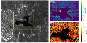
Recently, with the collaboration of Mario Marini, Margaux Bouzin, Laura Sironi, Laura D’Alfonso, Roberto Colombo, Giuseppe Chirico and Maddalena Collini, an infrared thermographic technique was tested (Bouzin et al. Nat. Comm. 2019), using a conventional thermocamera to reconstruct mesoscopic scale images (mm2-cm2) with a high spatial resolution (∼10 μm). The technique is based on the localization of temperature increases due to focused laser pulses and combines the temperature quantification with information on the thermic conducibility of the material, known the thickness and the adsorbed power. The technique was applied on a pipe organ (XVIII century) provided by the restorer Claudio Bonizzi (Ditta Inzoli Cav. Pacifico, Crema). The tin-based layer shows oxidation and alteration signs. The infrared analysis showed a correlation between the conducibility and the depth of the layer to the alteration state of the sample.